This year’s symposium introduced ‘the Untold stories of I&I’ and has shed light on some topics that are not discussed in detail in the regular curriculum of the I&I master. This symposium was held on 21 & 22 November 2013.
Speakers
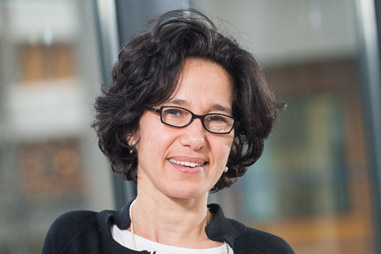
Dr. Maria Yazdanbakhsh – Human helminth infections and their bag of tricks
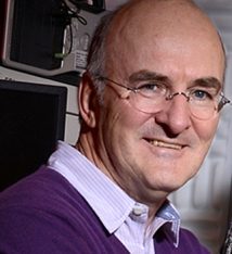
Dr. Michiel Pegtel – When a virus in control looses over its exosome
Dr. Peter Peters – Prion uptake in the gut: idication of the first replication sites
Eijkman seminar
Dr. Nicoletta Cieri – T memory stem cells at teh crossroad between protective and harmful immunity
Utrecht Life Sciences Seminar
Dr. Jan Liese – Intravital imaging of host-pathogen interactions during Staphylococcus aureus skin infection
Workshops
Dr. Peter Peters – The history and molecular mechanisms of prions
Dr. Nicoletta Cieri – Harnessing T cell immunity against cancer: tools, trials and tribulations
Leiden University Medical Center, Center of Infectious Diseases, Department of Parasitology, Leiden, The Netherlands
Human helminth infections and their bag of tricks.
Research synopsis-Maria Yazdanbakhsh heads the LeidenImmunoparasitology Group (LIPG). The group focuses on understanding how the immune system is shaped as a result of relentless challenge and modulation by pathogens in general, and parasites in particular. The team examines the immunological pathways at the population level by analysing responses ex vivoand in vitro. This allows the study of changes that take place in the immune system along the line of epidemiologic transition seen in many parts of the world and how this might result in epidemics of inflammatory diseases. Whereas both Th2 and Treg are hallmarks of the immune system in rural areas of the developing world, affluent countries are dominated by Th1 and Th17 responses. Combining population studies with in vitroand experimental assays the team has now identified single molecules that can condition dendritic cells to induce Th2 and Treg cells. Such molecules might have great potential in controlling inflammatory diseases.
Teaching/training/capacity building. Alongside regular curricular teaching, MariaYazdanbakhsh has placed much emphasis into training biomedical and medical students not only from the Netherlands and Europe but also from developing countries such as Indonesia, Ghana, Gabon and G. Bissau.
Part of the research activities of the group involves collaboration with centres in Indonesia, Gabon and Ghana. The long term capacity building, for over ten years, has been part of a strategy for collaborative groups to build up strong laboratory facilities and research teams.
Abstract:
Helminth infections have evolved with their human host and are masters of immune modulation. An important hallmark of helminth infections is the skewing of immune responses toward Th2. Population studies have shown increased intracellular IL-4 production and GATA-3 expression in CD4+ cells in areas where these parasites are highly endemic. Combining field studies in tropical areas where helminthes are endemic with molecular work in our laboratory in Leiden, it has been possible to show that helminths carry molecules that, in assays involving dendritic cell and T cell co cultures, lead to Th2 polarization. Characterization of one of these molecules, Omega-1 from Schistosomamansoni, has helped to dissect the signals that are needed for Th2 polarization. It isbecoming clear that TH2 responses can be involved in tissue repair and in glucose homeostasis. With respect to the latter, population studies have shown that in individuals carrying helminthes, the HOMA-IR is significantly improved, opening a new area for the use of immune modulators from helminth parasites in controlling insulin resistance.
VU University Medical Center Amsterdam, Department of Pathology, Exosomes Research group, Amsterdam, the Netherlands.
When a virus in control loses control over its exosomes
Michiel Pegtel completed his master’s degree from Groningen University. Next he moved to the National Cancer Institute (NCI/NIH) in Bethesda Md USA and obtained a PhD in virology/immunology at Tufts University medical school in Boston. Here he deciphered the role of the human tumor virus Epstein Barr in the pathogenesis of Nasopharyngeal Carcinoma by gene-expression analysis in collaboration with the Harvard/MIT Broad Institute. Next, Michiel performed post-doctoral research on cellular polarity and migration at the Netherlands Cancer Institute (NKI) in Amsterdam. For his work on functional transfer of viral microRNAs via exosomes, Michiel received the biannual Beijerinck Virology premium awarded by the Royal Dutch Academy of Sciences (KNAW). Michiel is currently assistant professor at department of pathology at the VU university medical center and heads the Exosomes Research Group, studying RNA transfer via extracellular vesicles in disease.
Abstract:
Innate detection of nucleic acids and type I interferons (IFNs) induction is fundamental in viral defense. Epstein-Barr virus (EBV) establishes a lifelong latent infection in more than 90% of human population. We study how latent EBV-infected B-lymphocytes communicate via exosomes with target cells in vitro and in vivo. By deep-sequencing we mapped the complete exosomal small RNA content and identified the incorporation of RNA polymerase-III-transcribed transcripts and demonstrate by gene-expression arrays and Elisa that EBV-RNA transfer via exosomes induces a potent IFN-mediated inflammatory responses. Systemic Lupus Erythematosus (SLE) patients have perturbed EBV control, increased viral loads and develop chronic inflammation in target organs. We show a link with EBV-exosomes and EBV that is consistent with viral RNA release and transfer in vivo. We propose that normal persistent latent herpes virus infection can potentiate autoimmunity in genetically predisposed individuals.
Netherlands Cancer Institute – Antoni van Leeuwenhoek, Division of Cell Biology, Amsterdam, the Netherlands.
Prion uptake in the gut: identification of the first replication sites
Prof. Peters obtained his PhD from Utrecht University, where he analyzed the ultrastructure of MHC class II antigen processing and discovered with Jacques Neefjes and Hidde Ploegh the ‘MHC class II compartment’ (MIIC) (Peters PJ et al., Nature, 1991, Peters PJ et al., J Exp Med 1995) and studied exocytosis of cytotoxic mediators in T cells. He established that secretory granules are of lysosomal nature (Peters PJ et al., J Exp Med. 1991). Peters joined the group of Rick Klausner at the National Institutes of Health in Bethesda, USA and identified ARF6 as a regulator for endocytosis (Peters PJ et al., J Cell Biol. 1995). In 1998, he became Principal Investigator at the Netherlands Cancer Institute and was appointed as professor at the Free University of Amsterdam. In 2010 he became partime professor of nanobiology at the Kavli Institute in Delft. Peters was the initiator to establish a 18 million Euro Netherlands Centre for Electron Nanoscopy that opened in October 2011 and is now part of the EU roadmap of large research infrastructure.
Summary of research interests.
One focal point of our structural biology group is to reveal and manipulate the macromolecular organization of cells under normal and pathogenic conditions at the nano-scale level. We use cryo-electron tomography of vitreous sections, currently the only method that can obtain molecular resolution of macromolecular machines in cells in a near-native situation. The tomograms contain a 3D map of the cellular proteome at about 3-4 nm resolution and we are just beginning to explore its potential by placing high priority on developing methods for nanotechnology. The other central point of our group is to visualise gene products in cells by electron microscopy at the highest resolution with gold probes on cryo-sections.
Abstract
Prions are infectious proteins composed of an abnormally folded isoform of the prion protein (PrPSc), the accumulation of which causes variant Creutzfeldt–Jakob disease, scrapie, and bovine spongiform encephalopathy, among other diseases. Prions propagate by converting endogenous, cellular prion protein (PrPC) into PrPSc containing a β-sheet core. Isolated PrPSccan be found in a wide range of aggregation states, from small oligomers to amyloid, and at least in larger aggregates the C-terminal portion of PrPSc acquires resistance to protease treatment. PrPC is a ubiquitously expressed protein that is most abundant in the nervous system. The accumulation of PrPSc causes morphological changes in the central nervous system including astrocytosis, neuronal cell loss and spongiform pathology and, in some types of prion disease, amyloid plaque formation. Pathology builds up during a long incubation period that ends in a short clinical phase and death. Expression of PrPC in the host is required for successful infection, since it provides the substrate for the conversion to PrPSc.
Peter will talk about how this successful infection takes place and what the major controversies where during the last 15 years of his collaborative effort with Noble Prize winner Stan Prusiner.
Interfaculty Institute of Microbiology and Infection Research Tübingen, University Hospital Tübingen, Germany
Intravital Iaging of Host-Pathogen Interactions during Staphylococcus aureus Skin Infection
| Curriculum Vitae | |
| since 2013 | Head, Hospital Hygiene and Infection Control, |
| Institute of Medical Microbiology and Hygiene, | |
| UniversityHospitalTübingen,Tübingen, | |
| Germany | |
| 2011-2013 | Research Physician, Institute of Medical Microbiology and Hygiene, |
| University Hospital Tübingen, Tübingen, Germany | |
| 2008-2011 | Research Fellow, Skirball Institute of Biomolecular Medicine, New York |
| University School of Medicine, New York, NY, U.S.A. | |
| 2008 | Board Certification for Microbiology, Virology, and Infection |
| Epidemiology | |
| 2002-2008 | Resident, Institute of Medical Microbiology and Hygiene, University |
| Medical Center Freiburg, Freiburg i. Br., Germany | |
| 1994-2001 | Study of Medicine, School of Medicine, Albert-Ludwigs-University |
| Freiburg, Freiburg i. Br., Germany |
Abstract
Advances in microscopy techniques over the last decades have contributed substantially to insights into the mechanisms employed by the host’s immune system to control and eliminate attacking pathogens. Intravital two-photon microscopy combines several technical advantages to allow for studying host-pathogen interactions on a single cell level in vivo, i.e. in a living organism.
Staphylococcus (S.) aureus is a clinically important pathogen, which is able to cause localinfections (e.g. in the skin and soft tissues) and severe generalized infections like sepsis and endocarditis. The ability of the host to control S. aureusis greatly dependent on the recruitment of neutrophils (PMN) to the site of infection.To gain more insight into PMN migration and host-pathogen interactions in vivo, we investigated a mouse model of S. aureusflank skin infection using intravital two-photon microscopy. For this purpose S. aureusreporter strains were generated and PMN migration in response to infection in vivowas then visualized using LysM-EGFP transgenic mice. PMN were rapidly recruited to the extravascular space of the dermis after injection of bacteria. After leaving the blood vessels, the cells exhibited directed movement towards the focus of infection. Interaction with S. aureusinduced a change in the migratory pattern of the PMN, which was characterized by decreased velocity and track straightness. Furthermore, depletion of neutrophils or blocking G-protein coupled receptors (GPCR) lead to an uncontrolled proliferation of bacteria. Tracking of transferred labeled bone-marrow derived neutrophils showed that PMN recruitment to the site of infection is dependent on GPCR on the neutrophils themselves, whereas IL-1-receptor was required on host cells other than PMN.Overall, two-photon microscopy is a powerful tool to investigate the dynamics of the immune response, bacterial cell location, and gene expression in vivoon a single cell level during S.aureus infections.
Experimental Hematology Unit, San Raffaele Scientific Institute, Milan, Italy
T memory Stem Cells at the crossroad between protective and harmful immunity
| Curriculum Vitae | |
| 2011 – present | Vita-Salute San Raffaele University Milan
PhD research (PhD program in Molecular and Cellular Biology), investigating the functional role of human memory stem T cells in health and disease. |
| March 2009 – July 2010 | San Raffaele Scientific Institute Milan
Thesis internship in the laboratory of Experimental Hematology of Dr. Bonini, investigating the plasticity of T cells in the context of adoptive immunotherapy of cancer. |
| September 2006 – September 2007 | San Raffaele Scientific Institute Milan
Internship in the laboratory of Molecular and Cellular Neurobiology of Dr. Meldolesi, investigating the role of TREM- 2 in microglia biology. |
| September 2005 | San Raffaele Scientific Institute Milan
One month internship in the laboratory of molecular Genetics of Kidney Diseases of Dr. Rampoldi, covering the basic techniques of cell biology and biochemistry. |
In 2013 Nicoletta Cieri won 2013 the Jon J. Van Rood Award at the 39th annual European Bone Marrow Transplantation (EBMT) Congress in London, Great Britain.
Abstract:
The ability to remember and respond more robustly in a second encounter with a pathogen is a pivotal property of the adaptive immune system and forms the basis of vaccination and adoptive cellular therapy of cancer. This process has been proposed to involve a stem cell-like memory T-cell subset, able to rapidly differentiate and self-renew upon antigen re-encounter. The characterization of such a T-cell subset is not only of basic interest but also of clinical relevance for the development of strategies to target pathogens and cancer by adoptive T-cell therapy. Such a memory T-cell subset, referred to as memory stem T cells (TSCM), has been recently described in humans (Gattinoni et al., 2011).
Our group identified conditions able to instruct naïve T cells into TSCMcells in vitro, thus allowing their expansion and genetic modification in clinically compliant conditions (Cieri et al., 2013). Gene-modified TSCM, defined as postmitotic CD45RA+ CD62L+ CCR7+ IL-7Ra+ CD95+ T lymphocytes, are endowed with exceptional persistence and functional capacity invitro andin vivo, which could be exploited for cancer adoptive immune-gene therapy.Nevertheless, while self-renewing TSCMwould be highly effective in providing long-term immune-surveillance against pathogens and cancer cells, this very same cell subpopulation may also represent a foe when considering T-cell mediated pathologies, such as autoimmune diseases and graft versus host disease (GvHD). In these clinically relevant contexts, TSCMmay represent a reservoir of long-lived T cells with undesired and detrimental specificities responsible for therapy resistance and high morbidity. Indeed, we have documented a selective accumulation of TSCMearly after allogeneic hematopoietic stem cell transplantation (HSCT), and our data on a small cohort of transplanted patients suggest that the extent of TSCMaccumulation one month after HSCT correlates with GvHD occurrence and severity. Of note, we found higher TSCMfrequencies also in patients with active rheumatoid arthritis, corroborating the notion that TSCMmay represent the latent reservoir of lymphocytes responsible for disease perpetuation. Thus, TSCMcells represent a double-edged sword that should be exploited in adoptive immunotherapy, but tamed in the context of T-cell mediated disorders.
Here, I will review recent advances in the comprehension of TSCMbiology and ontogeny and how this knowledge may guide the development of strategies directed at selectively hamper or expand this cell population in selected clinical conditions.


